It’s a lazy, sunny Saturday afternoon. The most important thing on your list for the day is playing with the dog. You throw the frisbee, Buddy jumps into the air to catch it and falls to the ground. He gets back up on his feet, but he has a limp.
Of course he has a limp! He just hit the ground pretty hard. But a few days go by and the limp is still present. So, you load him up and drive to your local veterinarian. After examining Buddy, your veterinarian informs you that Buddy is suffering from a torn cranial cruciate ligament.
So where do you go from here?
Will you ever get to throw the frisbee with your furry friend again?
Will he ever walk without a limp?
Cranial Cruciate Ligament Injuries: The Basic Facts
To better understand the condition and process of correction, let’s lay out the basics.
Much like humans’ anterior cruciate ligament (ACL), a dog’s cranial cruciate ligament (CrCL) acts as the main support for the knee joint (stifle).

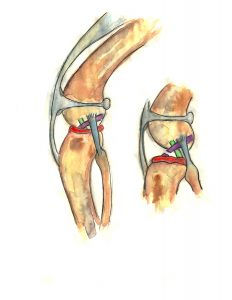
Figure 1: Comparison of normal cruciate (first image) to a torn cranial cruciate ligament (second image)
Red: meniscus; Purple: cranial cruciate ligament; Green: caudal cruciate
As shown in the picture above, the meniscus lies between the femur and tibia bones and acts as a cushion. When the CrCL ruptures, the meniscus suffers damage as well.
Cranial cruciate ligament disease (CrCLD) is one of the leading causes of hind limb lameness in canines. The tear of the CrCL can be partial or complete. However, a partial tear will most certainly become a complete tear if the condition is not corrected. Cruciate tears, partial or complete, will cause pain and eventually arthritis.
It is rare, but very possible, for a cruciate ligament tear to result from acute injury. More often, the ligament degenerates over a period of time.
Are Certain Dogs Predisposed to CrCLD?
Big or small, old or young, any dog can tear the CrCL. It is very rare in cats. However, studies have shown that certain factors do play a role in this disease.
- Aging of the ligament
- Obesity
- Genetics
- Breed
- Physical condition
- Body conformation.
Studies have also shown that 40-60% of dogs who tear one CrCL will tear the other one at some point in time.
Unfortunately, keeping your dog in the best shape is the only way of prevention.
Signs/Symptoms of CrCLD
- Limping (severity varies)
- Stiffness
- Reluctance to jumping/playing
- Difficulty rising from sit/lay position
- Swelling or pain around the stifle
- Muscle atrophy
- Pain
- Popping sounds from the stifle.
What Are My Treatment Options?
If found in time, a CrCL that is partially torn may be corrected with prolotherapy. This should only be considered on a case to case basis, as it is not always the best choice.
Surgery is the recommended treatment. Furthermore, stabilizing the joint may only be achieved via surgery. Until the joint becomes stable, the pain will continue. There are several different methods of surgery, including:
- Tibial Plateau Leveling Osteotomy (TPLO)
- Tibial Tuberosity Advancement (TTA)
- Suture-based Techniques
- Extra-capsular Suture Stabilization
- Tightrope®.
All three methods have different approaches and, of course, different costs. According to recent studies, however, all of these methods have comparable outcomes approximately one-year post-surgery. The differences may be discussed with your veterinarian, as some methods require special training.
The Extra-Capsular Suture Stabilization Method
Here at White Oak Animal Hospital, we most commonly use the extra-capsular suture stabilization method, which is what we will cover in this article.
This procedure may also be referred to as the lateral suture method, ex-cap suture method, lateral fabellar suture stabilization or the fishing line technique.
Figure 2: Representation of Extra-capsular Suture Stabilization Method
As illustrated in the picture above, this method requires that a heavy, sterile suture be placed on the outside of the joint. The suture is positioned in a way to mimic the action of the CrCL. Over time, this method causes a buildup of scar tissue around the stifle joint, which provides stability for when, or if, the suture deteriorates.
As mentioned above, all possibilities should be discussed with your veterinarian to find the best method for your dog.
Are There Options Other Than Surgery?
Yes. Sometimes there are cases not fit for surgery! The non-surgical treatments are:
- Pain medicine/supplements
- Restricted activity
- Rehabilitation therapy
- Custom knee braces.
Great care should be taken with pain medicine. Long-term use may cause severe liver/kidney damage. Routine blood work may be required.
Custom knee braces are new to the veterinary world. Because of this, there is a lack of evidence.
Aftercare
Postoperative care is just as important as the surgery itself! No matter how great your pup seems to feel after surgery, he/she must be under restricted activity for several weeks after surgery. This could mean kenneling your dog when unsupervised and leash-walking for bathroom breaks.
Rehabilitation therapy speeds recovery AND improves the final outcome.
It is recommended to keep your furry friend on a glucosamine supplement as arthritis is very likely to develop, with or without surgery. The physical condition of your pup, including weight, is very important to keep in check!
In most cases 85-90% of the dogs undergoing CrCL repair improve significantly as long as they get appropriate post-surgical care.
Powerful Tools to Help Your Dog’s Ligament Challenges
There are many quick and easy changes you can make at home to help you give your dog an edge on easing tendon and ligament challenges.
- Learn more about torn ligaments and cruciate disease.
- Provide joint support. PET | TAO Harmonize Joint Supplement is a blend of Eastern herbs and Western nutraceuticals working together to lubricate and restore your dog’s joints.
- Ease your dog’s discomfort naturally. PET | TAO Comfort is a blend of Eastern herbs and Western supplements to soothe your dog’s arthritic challenges to make him/her more comfortable.
- Try PET | TAO Freeze Dried Beef Liver Treats. According to TCVM, liver controls tendons and ligaments. As few as 5-6 treats per day can make a huge difference in your dog’s tendon and ligament health!
- Try a Blood-building TCVM Diet. PET | TAO Zing Canned Formula builds Blood. According to TCVM, Blood deficiency leads to ligament tears.
- Learn more about TCVM Herbal Remedies. Chinese medicine offers many amazing natural solutions for ligament and cruciate challenges. Some good examples are:
Related Posts
-
Prolotherapy Sessions for Cranial Cruciate Ligament (CCL) Injuries
Will prolotherapy sessions help my pet? Possibly! Prolotherapy sessions help many pets and take the…


